Knee Replacements

Knee replacements is a common orthopaedic procedure in Australian hospitals. In Australia, 62,800 knee replacements were performed in 2020-21, compared to 38,800 hip replacements


What Exactly Is A Knee Replacement?

A knee replacement, otherwise known as a knee arthroplasty, is a surgical procedure to replace either part of or the whole of the knee joint. Performed by an Orthopaedic Surgeon, these surgeries hospital stay of a week or so.


How Common Are Knee Replacement Surgeries?

Knee replacements is a common orthopaedic procedure in Australian hospitals. In Australia, 62,800 knee replacements were performed in 2020-21, compared to 38,800 hip replacements (AIHW, 2023).


What Are The Reasons For A Knee Replacement?

There are multiple reasons for needing a replacement… whether it’s due to degenerative changes, traumatic injuries or as a measure to help people manage better with less pain in their knee. A common group we see though are older adults who are having increasing issues with Osteoarthritis (OA) in their knees.

In Australia, 1 in 5 adults over the age of 45 have Osteoarthritis
— AIHW, 2023

Knee Osteoarthritis can be divided into two categories

Primary Knee OA which refers to degenerative changes in the articular cartilage which can’t be attributed to a particular cause though changes become more common as we progress through the lifespan

Secondary Knee OA refers to degenerative changes that can be attributed to a particular cause. These causes my include obesity, prior traumatic injury, immobilisation, joint instability / hypermobility, and Vitamin D deficiency

It is important to keep in mind that any changes in your knees don’t necessarily lead to a joint replacement. In fact the Royal Australian College of GP’s Guideline for managing Knee OA recommends referral to a surgeon ONLY when conservative options have failed for end-stage OA.


Functional Knee Anatomy

The knee is the largest joint in the body. Not just one joint, but consisting of three seperate joints!

The bones involved in making up the knee are;

  • Tibia or your shin bone and the top section provides a platform for your thigh to rest on.

  • Patella or the kneecap acts as a fulcrum to transfer the force of your thigh muscles on to the Tibia.

  • Femur or the thigh bone and is also the largest long bone in your body. The bottom of this bone rests on top of the Tibia.

  • Fibula which runs down the outside of your Tibia

Yes, There Are Three Knee Joints!

Tibiofemoral (Tibia & Femur) Joint: This joint is classified as a ‘Modified Hinge’ joint, and its primary motions are flexion / extension and external rotation of the Tibia.

Patellofemoral (Patella & Femur) Joint: It is classified as a “Saddle” joint, and the primary motions are gliding up, down, and side to side relative to the tibiofemoral joint.

Superior Tibiofibular (Tibia & Femur) Joint: This is the joint located at the top of the Tibia and Fibula and it is classified as a 'Plane” joint. The primary motions here are to allow the two bones to move forwards / backwards, up / down, and rotate in relation to each other.


To make things more confusing, the bones and joints are grouped in to 3 compartments.

Lateral Compartment (Outside Part Of The Knee)

Consisting of the lateral end of the Femur and the lateral end of the top of the Tibia.

Medial Compartment (Inside Part Of The Knee)

Consisting of the medial end of the Femur & the medial top of the Tibia.


Patellofemoral Compartment (Front Of The Knee)

Consisting of the Patella and the end of the Femur.


What Are The Treatment Options For Knee OA?

Historically knee surgeries such as a total knee replacement or an arthroscope were one of the first lines of treatment in knee OA. As time has gone on however, the focus on surgery has diminished, instead shifting towards education, exercise & pain relief as needed.


First Treatments

Education, Exercise & Weight advice are indicated for all people experiencing knee pain from OA


Second Treatments

This is the introduction of strategies to help you with your symptoms. This might be medicine, bracing or passive therapies but is only required in some cases


Third Or Last Treatment Options

This includes surgeries but are only indicated in severe cases and should only be utilised when the other treatments are no-longer helpful


Conservative Management

In individuals with Knee OA, conservative management with a Physiotherapist is typically the first line treatment. The aim of this is to address the restrictions in knee range of motion, pain, strength and functional limitations. The symptom in particular conservative management aims to address is knee pain, as the associated reduction in functional ability strongly relates to future disability and dependency. As a result, early treatment of the associated pain related functional decline is vital for treating OA & other forms of knee pain (Farrokhi et al; 2016).


Strength training

Strength training is one of the core components for conservative management of knee OA. Exercises generally focus on lower limb strengthening, but can include upper limb exercises as well.

According to Vincent et al; (2012), the basic components of a lower limb strengthening program for knee OA include;

  • Squatting type exercises

  • Knee Extension

  • Knee Flexion

Other common exercises that are commonly prescribed in the conservative management of knee OA target hip abduction/ adduction, and ankle strength. The reasoning behind this is addressing any potential contributing factors to altered kinematics of the knee (Vincent et al; 2012).


GLA:D Protocol

One of the most popular lower limb strengthening strategies utilised by physiotherapists is the Good Living with Osteoarthritis in Denmark, or GLA:D, program. It has been demonstrated to improve pain intensity and quality of life for people with osteoarthritis (Skou et al; 2017). The GLA:D program is a structured exercise & education program, with guidelines around exercise prescription, dosage, and education surrounding the prognosis of OA.

Education around the pathology of osteoarthritis, prognosis, and exercise almost always form a major part of conservative management. This forms a part of the GLA:D protocol, with 8 weeks of education a core component of it.


Knee Replacement Surgery - When All Else Fails

If all conservative management strategies for knee OA fail to improve the quality of life for the individual to an adequate level, then surgical interventions is the next port of call.

Osteoarthritis is the most common condition leading to both hip and knee replacements, with this trend increasing by on average 2.6% per year between 2010-11 & 2020-21 (AIHW, 2023).

Knee replacement surgery can offer good outcomes for patients who require these surgeries, but it is important to discuss with your surgeon the risks & benefits of each procedure to ensure your expectations are in line with each other.

Main types of knee replacement

There are two main types of knee replacements. The first one is a Partial knee replacement, otherwise known as a Unicompartmental knee placement. The second one is a Total knee replacement, also known as a total knee arthroplasty.

In a Partial knee replacement (UKR) only one portion of the thigh bone (femur) is replaced, along with the corresponding portion of the shin bone (tibia) opposite to it. This could be the medial compartment, or the lateral compartment.This can be seen on the left sided diagram on Image 1 below.

In a Total knee replacement (TKR) the entire distal (or furthest) portion of the femur, and the proximal (or nearest) portion of the tibia is replaced. Essentially, all three compartments of the knee joint are replaced. As mentioned above, this is also known as a total knee arthroplasty. An example of this is shown on the right sided diagram below.

TKR vs UKR

Generally, a UKR offers a shorter hospital stay and rehabilitation compared to a TKR (Pongcharoen et al; 2023). According to the Australian Orthopaedic Association (2015), 88% of knee replacement procedures are TKR’s, with UKR’s making up 5%, and revisions of previous procedures making up the remaining 7%.

UKRTKR
DURABILITYRoughly 10% have to be replaced in around 10 yearsRoughly 5% have to be replaced in around 10 years
REHABILITATIONRehabilitation usually only involves outpatient physioUncomplicated TKR's can rehab at home
RETURN TO FUNCTIONAround six weeks to return to most activities3 - 6 months to fully return to most activities
SATISFACTIONAround 90% of UKR patients would undergo the procedure againAround 80% of TKR patients would undergo the procedure again
RISK OF COMMPLICATIONS3% experience complications in the first 12 months5% experience complications in the first 12 months

Comparison of TKR Vs UKR (Informed Health, 2006)


Following the surgery

What will I experience initially?

Initially, individuals who undergo either a UKR or a TKR experience swelling, pain, and restricted knee range of motion. The main goal in hospital post surgery is to get you ready to return home in a safe manner as quickly as possible.

Swelling is usually quite severe initially, lasting for the first few weeks. Up to six months post surgery, mild to moderate swelling may persist. Swelling usually peaks in intensity 7-10 days post surgery.

Pain is often severe post total knee replacement. As a result, individuals are often prescribed strong painkillers (i.e. Endone) as a part of the initial management of symptoms.

Knee range of motion is usually addressed in a progressive nature with the input of a physiotherapist.

What comes next?

Post knee replacement, rehabilitation often commences on the same day. Typically TKR rehabilitation protocols occur for a longer duration, compared to a UKR. but the principles of rehabilitation are similar between both.

Weeks 0-3- Initial stages

This can involve a simple range of motion exercises in surrounding joints, and weight bearing (depending on surgeon's orders) on the affected side. Individuals who have undergone a UKR can commence more advanced resistance training.

Common Exercises: Ankle Pumps, Heel slides, Inner Range Quads, Passive knee straightening

Functional Training: Lying to Sitting, Sitting to Standing, Mobilising as tolerated (depending on surgeons orders)

Goals: Control pain, Improve swelling

Weeks 4-6- Intermediate stage

This stage involves progressive exercises compared to the previous stage. Individuals who have undergone a UKR can commence more advanced functional training earlier than TKR.

Common Exercises: Active range of motion, Heel raises

Functional Training: Normalising walking with a stick & progression to a single point stick

Goals: Knee flexion >90 degrees

Weeks 7 - 12- Advanced stage

This stage involves more functional progression, with an addition of resistance training & compound exercises (i.e. squats)

Common Exercises: Squats, Resisted knee range of motion (hamstrings, quads, adductors, gluteals)

Functional Training: Exercise bike, Supervised walking with a stick

Goals: Knee ROM 0 degrees extension - >110 degrees flexion

3+ Months- Maintenance

This stage is very patient specific. For more information on why, please read the section below.

Common Exercises: Exercise bike (increased distance), Return to gentle sports, Return to gentle gym workouts

Goals: Continue exercise program to optimise patient outcome from surgery

How long will it take to regain my function?

This is a complicated question, and it depends on your long term goals.

It is important to emphasise that a knee replacement will not allow you to engage in physical activity that you were unable to do prior to the surgery. Knee replacements are not designed to allow an individual to re-engage in high impact activities (AAHKS, 2017). The ability to engage in unlimited low impact activities, such as walking, golf, swimming, cycling etc. is a realistic goal.

Generally, it can take up to a year to recover fully from a total knee replacement, while it can take up to 4 months to recover fully from a unilateral knee replacement.

Wrap up

Knee pain contributes to a significant burden in Quality of life in many individuals. One of the chief contributing factors of knee pain is OA, particularly in individuals over the age of 45. Conservative management is typically the recommendation for people experiencing knee OA symptoms, with surgical interventions not indicated until conservative and symptomatic management fails to be enough. Physiotherapy led exercise programs like GLA:D have become increasingly popular due to the effectiveness of the intervention.

Rehabilitation post knee replacement generally consists of progressive range of motion, strengthening, and functional training. The duration of rehabilitation varies, depending on the nuanced presentation of your complaint and the type of surgery carried out. Whatever the case, an experienced physiotherapist will be able to work with you to help you regain your function again and maximise your outcomes following your surgery.


References

Australian Orthopaedic Association National Joint Replacement Registry. Annual Report. Adelaide:AOA; 2015. Available at: https://aoanjrr.sahmri.com/documents/10180/217745/Hip+and+Knee+Arthroplasty.pdf/02ee14ed-39a7-46cd-9589-03728238e475?version=1.1&t=1443634264710&download=false (Accessed: 15 December 2023).

Buhagiar, M. A., Naylor, J. M., Harris, I. A., Xuan, W., Kohler, F., Wright, R., & Fortunato, R. (2017). Effect of inpatient rehabilitation vs a monitored home-based program on mobility in patients with total knee arthroplasty: the HIHO randomized clinical trial. Jama, 317(10), 1037-1046.

Chronic musculoskeletal conditions : Osteoarthritis (no date) Australian Institute of Health and Welfare. Available at: https://www.aihw.gov.au/reports/chronic-musculoskeletal-conditions/osteoarthritis (Accessed: 21 October 2023).

Full vs Partial | Hip and Knee Care. (n.d.). AAHKS Hip and Knee Care. https://hipknee.aahks.org/full-vs-partial-knee-replacement-whats-the-difference/

Information, N. C. for B., Pike, U. S. N. L. of M. 8600 R., MD, B., & Usa, 20894. (2019). Osteoarthritis of the knee: What different types of knee implants are there? In www.ncbi.nlm.nih.gov. Institute for Quality and Efficiency in Health Care (IQWiG). https://www.ncbi.nlm.nih.gov/books/NBK544988/

Pongcharoen, B., Liengwattanakol, P., & Boontanapibul, K. (2023). Comparison of Functional Recovery Between Unicompartmental and Total Knee Arthroplasty: A Randomized Controlled Trial. The Journal of Bone and Joint Surgery. American Volume, 105(3), 191–201. https://doi.org/10.2106/JBJS.21.00950

RACGP - Guideline for the management of knee and hip osteoarthritis. (2018). Racgp.org.au. https://www.racgp.org.au/clinical-resources/clinical-guidelines/key-racgp-guidelines/view-all-racgp-guidelines/knee-and-hip-osteoarthritis

Skou, S. T., & Roos, E. M. (2017). Good Life with osteoArthritis in Denmark (GLA:D): evidence-based education and supervised neuromuscular exercise delivered by certified physiotherapists nationwide. BMC Musculoskeletal Disorders, 18(1). https://doi.org/10.1186/s12891-017-1439-y

Vincent, K. R., & Vincent, H. K. (2012). Resistance exercise for knee osteoarthritis. PM & R : The Journal of Injury, Function, and Rehabilitation, 4(5 Suppl), S45-52. https://doi.org/10.1016/j.pmrj.2012.01.019

Read More
Condition Summary, Physiotherapy, Shoulder Jesse Dean Condition Summary, Physiotherapy, Shoulder Jesse Dean

Shoulder Dislocations & Their Management

Shoulder Dislocations Are A Common Sporting Injury But What Is It Exactly & How Does A Surgical Repair Compare With Conservative Treatment?
This past month football fans may have seen a few devastating injuries and this is the unfortunate part of sport at this level. One of which was the shoulder dislocation injury to St. Kilda young tall forward Max King, of which was a recurrence of the same injury that required surgical intervention only last year. Over the weekend that he took to the field less than a month post injury that was initially deemed to be season ending.

Understanding Shoulder Dislocations Picture your shoulder joint as a ball and socket like a golf ball sitting in a golf tee, where the upper part of your arm bone (humerus) fits into a shallow socket in your shoulder blade (scapula). This joint allows you to move your arm in many directions, giving you the ability to lift, rotate, and swing your arm freely. Unfortunately a larger range of movement is at the detriment of stability. (Belk et al. 2023)

Now, a shoulder dislocation occurs when the ball part of the joint (the humerus) slips out of its socket in the shoulder blade. This can happen due to various reasons, such as a sudden impact, a fall, or a forceful movement of the arm. When this happens, it can be quite painful and restrict your ability to move your arm normally.


There are two main types of shoulder dislocations:

Anterior Dislocation

Posterior Dislocation

This is the most common type, where the humerus moves forward and out of the shoulder socket. You might notice a visible bulge in the front of your shoulder, and you may be unable to move your arm.

The extreme forces generated during tackles, falls, and collisions in sports like Football, Rugby, Netball & Basketball can lead to shoulder instability and dislocation, often causing severe pain and hindering functional ability. With some severe injuries sustains bony injuries such as Hill-Sachs or Bankart type dislocations that have fractures of the socket in conjunction with soft tissue tearing/straining (Watson et al. 2016)

These injuries highlight the need for comprehensive medical attention, proper rehabilitation, and preventive measures to ensure a safe and successful return to the game.

Additionally, it underlines the significance of educating players, coaches, and support staff about shoulder injury prevention and management strategies.

Less common, this type happens when the humerus moves backward out of the socket usually as a result of falling on your arm as it is outstretched. It might not be as obvious as an anterior dislocation, but you'll still experience pain and limited arm movement (Handoll & Al-Maiyah, 2004).


Preventive Measures For Athletes Across All Levels

Pre-Season Screening & Assessment

Conducting thorough pre-season screenings can help identify players with pre-existing shoulder issues or risk factors for dislocations. A comprehensive assessment of shoulder strength, flexibility, and stability can aid in designing individualised training programs to strengthen weak areas and reduce vulnerability to injuries.


Shoulder Strengthening & Stability Training

Following on from above implementing targeted shoulder strengthening exercises in athletes conditioning routines is crucial for injury prevention. Focusing on the rotator cuff muscles and surrounding shoulder stabilisers can enhance joint stability and reduce the incidence of dislocations.


Proper Technique & Sport Specific Drills

Coaches should emphasise teaching players proper position & sport specific techniques to minimise the risk of shoulder dislocations. By focusing on form, players can avoid putting excessive strain on their shoulders during physical encounters. Incorporating controlled drills and practicing safe landings can also help in reducing injury risks.


Warm-Up & Proper Preparation

Encouraging players to adhere to effective warm-up routines is essential in preparing the shoulder joint for physical activity. Dynamic stretching, light resistance exercise and joint mobility exercises can improve proprioception and flexibility, reducing the likelihood of injuries like shoulder dislocations.


Injury Management & Rehabilitation

If a player does suffer a shoulder dislocation, prompt and appropriate medical attention is crucial. The sooner the athlete has the joint reduced back into the joint the less time the joint soft tissue is stretched and therefore better outcomes long term. An accurate diagnosis, followed by a comprehensive rehabilitation program, can aid in a smoother recovery process and reduce the risk of recurring dislocations.


Once an MRI supported dislocation has been identified the next decision is how best to treat it, when it comes to treating shoulder dislocations, healthcare professionals often face the decision between operative and non-operative approaches.

While both methods aim to restore joint stability, each has its own set of advantages and disadvantages.

Operative treatment involves a specifically matched surgical procedure to repair the damaged structures within the shoulder joint. The most common surgical technique is known as an internal fixation. This procedure entails reducing the dislocated shoulder joint to its normal position and using various surgical techniques like sutures, anchors, or screws to stabilise the joint.


Positives of Operative TreatmenT

Immediate Stability

Surgical intervention provides immediate stabilisation of the dislocated shoulder joint, reducing the risk of recurrent dislocations.

Reduced Recurrence Rate

Several studies have shown that operative treatment significantly lowers the recurrence rate of shoulder dislocations compared to non-operative methods.


Negatives of Operative Treatment

Surgical Risks

Like any surgical procedure, there are known risks, including infection, nerve damage, and blood vessel injury.

Longer Recovery Time

Post-operative rehabilitation and recovery can take longer compared to non-operative treatment, requiring patience and adherence to a rehabilitation program.

Prolonged Immobilisation

Prolonged use of a sling or brace can lead to stiffness and muscle atrophy, potentially delaying the return to normal activities.

Non-operative treatment typically involves a process known as "closed reduction," where a healthcare professional manually manoeuvres the dislocated shoulder back into its socket. Following reduction, the patient undergoes a period of immobilisation using a sling or shoulder brace. Subsequently, physiotherapy and exercises are prescribed to regain shoulder strength and stability, especially overhead or out to the side (Watson et al. 2016).

Positives Of Non-Operative Treatment

Avoidance Of Surgical Risks

Non-operative treatment avoids the potential risks associated with surgery, making it a safer option for certain patients.

Lower Healthcare Costs

Compared to surgical intervention, non-operative treatment tends to be far more cost-effective.


Negatives of Non-Operative Treatment

Severe dislocations have Increased Recurrence Risk

Studies have indicated a higher recurrence rate for non-operative treatment for severe dislocations, especially in younger, active individuals.

While both operative and non-operative treatment have been shown to be effective the reality is some athletes of all levels don’t return to the activity they were doing pre-injury. Van Lersel & colleagues 2023, conclude the largest factor contributing to this is fear of re-injury with 20% of participants stating this to be their main reason, with functional demands just behind, both of which we aim to address with shoulder rehab programs.


Read More

Joint Hypermoblity Syndrome

IS FLEXIBILITY ALWAYS A GOOD THING?

As someone with hypermobilty himself, Luke (physio) discusses the spectrum of mobility and what the implications of being too stretchy can mean.

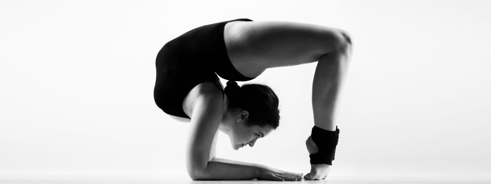
If you’ve always thought of yourself as double jointed or have always strived to achieve the level of mobility that a contortionist posses, this might the article for you!

Formally known as Benign Joint Hypermobility Syndrome (BJHS), Joint Hypermobility Syndrome (JHS) is a heritable disorder of the connective tissues. People typically present with very elastic skin, a large amount of flexibility and often with a history of joint injuries.

 
Luke Anthony - Physio
I’ve always found stretching to be quite easy and I’ve always been surprised when my friends couldn’t touch their toes. Yoga has always been easy for me, but little did I know that it was because I was hypermobile rather than a Yogic Legend.
It wasn’t until I had some joint dislocations & other weird musculoskeletal conditions that I was screened for hypermobility. Thankfully my GP referred me to a rheumatologist & to have my heart checked as hypermobility can affect a lot of different structures throughout the body!
— Luke Anthony - Physiotherapist

What Is The Prevalence Of Hypermobility

  • Affects 10-20% of the population in Western countries and higher still in Indian, Chinese, & Middle Eastern populations

  • More common in children and decreases with age

  • Hypermobility is more common in women than men

  • There are wide variations between ethnic groups

What Are The Causes Of Hypermobility?

JHS is commonly found to have a hereditary factor, in particular a genetic variation in collagen. Collagen is found throughout the body in structures such as the skin, ligaments, and the heart valves. If collagen is weaker than it should be, there can be increased laxity in these structures which can lead to issues.


Does It Really Matter If I’m Stretchy?

We believe it is important to screen patients for hypermobility that could be contributing to their symptoms. Ross and Grahame (2011) describe the following factors that highlight this importance:

  • Inappropriate and potentially harmful labelling of treatments may be applied on the basis of an erroneous diagnosis such as rheumatoid arthritis, hypochondriasis, or somatisation.

  • Over zealous physical manipulation causing avoidable damage, such as dislocations, connective tissue ruptures, or fracture of fragile bone.

  • Exercise therapy may be excessively forceful or ineffectual.

  • Anecdotal evidence exists that orthopaedic operations on patients with connective tissue disorders, leading to poorer outcomes.

  • Chronic pain may sometimes lead to a potentially reversible downward spiral or immobility, reconditioning, dependency, and despair.


What Are Some Of The Clues Suggesting Joint Hypermobility?

Hypermobility In Children & Adolescents

unsplash-image-tJHU4mGSLz4.jpg
  • Coincidental congenital dislocation of the hip

  • Late walking, with bottom shuffling instead of crawling

  • Recurrent ankle sprains

  • Poor ball catching and handwriting skills

  • Tiring easily compared with peers

  • So called growing pains of chronic widespread pain

  • Joint dislocations

Hypermobility In Adults

unsplash-image-5H0p6JPUHbI.jpg
  • Non-inflammatory joint or spinal pain

  • Joint dislocations

  • Multiple soft tissue injuries

  • Increase in pain or progressive intensification of pain that is largely unresponsive to pain medications

  • Progressive loss of mobility owing to pain or kinesiophobia (pain avoidance through movement avoidance)

  • Premature osteoarthritis

  • Autonomic dysfunction such as dizziness or faintness

  • Functional gastrointestinal disorders (sluggish bowel, bloating, rectal evacuator dysfunction)

  • Laxity in other supporting tissues such as hernias, varicose veins, or uterine or rectal prolapse


How is Hypermobility Diagnosed In Physiotherapy?

A huge part of a physiotherapy assessment is the subjective assessment, the interview your physio does with you at the start of a session. It helps us to learn about your injury, family history, previous problems etc… If we consider some of the clues from the previous section, a thorough subjective assessment can help build a Physio’s suspicion that hyper mobility may be a factor in your complaint.

When looking at things that can be tested to confirm a suspicion of hypermobilty, a physiotherapist will typically use a screening test called the Beightons Score. This test assesses the amount of movement in your elbows, knees, hands & trunk. Scored out of 9, with a score of > 4 typically considered to be a positive finding for hypermobility (Clinch et al. 2011). Though the Beightons Score is a great tool to quickly give an impression of a patients mobility, it does have limitations as it doesn’t look at the wider system such as the shoulder, hip or ankle.

Beightons Score

If your physiotherapist holds concerns about your level of hypermobility, they may refer you back to your GP who may request blood tests and / or refer you to a Rheumatologist for a more thorough investigation.


How Can Physiotherapy Help?

The role of a physiotherapy can vary greatly depending on the presentation of a patient. The team at Richmond Rehab are passionate about having a positive influence in the areas we can help with, and directing patients to a more appropriate service when required. We believe that simply identifying patients with JHS and educating them about how it can influence their symptoms is an essential step in helping a patient manage their symptoms. When a patient has a good understanding, they then have the power to modify their behaviours and rely on passive treatment less.

Depending on the severity of presentation, your physiotherapist may opt for some supportive taping in the short term and possibly bracing for long-term management if required.

The one thing that all Physio’s should do however, is to develop an exercise program suitable for your specific presentation. Palmer et al. (2013) reports that exercise interventions can create improvements with JHS over time without any adverse effects. The mode of exercise was inconclusive however, with generalised exercise performing better than joint-specific exercise and that further studies in the area are indicated.

Is Massage Or Manipulation Helpful?

The difficulty for patients in pain with undiagnosed JHS is that they are not best placed to decide which treatments are best suited for them. Typically looking for a relief in pain, they often bounce from therapist to therapist seeking short-term relief from passive treatments.

In relation to helping to ease the pain, manual therapies can have their place, however they do little to create any longterm changes. Instead they can create a reliance on practitioners to help ease a patients pain and potentially lead to a lot of costly appointments over time.

We believe it is imperative to identify hypermobile patients as early as possible to allow us to develop an appropriate management plan that is guided by evidence. We may refer JHS patient for manual therapy if they require temporary relief, however our focus is always on education and self-management in the longterm.


So What Are We Saying?

The purpose of this article is to illustrate to you that more mobility isn’t necessarily a good thing. Sure it’s great to have a good level of mobility to move well, but there are limits. If you’ve always thought of yourself as being quite bendy and have been struggling with ongoing musculoskeletal pain, it might be time you had a chat with your physio or GP.

We’d be thrilled to see more patients educated about their bodies and taking a proactive approach to managing their health. When you’re informed you can make better decisions about which treatments are best for you and how you’d like to spend your hard earned money.


Read More

Acute Lower Back Pain & Myotherapy

Lower back pain (LBP) is one of the world’s leading musculoskeletal complaints. There is a high chance you’ve experienced it and here are some tips on what you can do to manage it.

There is a likely chance that you will experience some form of lower back pain (LBP) in your life as LBP is one of the world’s leading musculoskeletal complaints, and most LBP is acute; meaning that it may persist up to a period of 12 weeks, the first occurrence usually happening between the ages of 20-40 years old (Casazza, B.A., et al).

Now, there is always the chance that your LBP may resolve on its own over the next few days to few weeks, but it is a good idea to ask for help and advice if the pain does not improve or worsens over the next few weeks, or if the pain stops you from performing your everyday activities.

Back Pain.jpg

Who is at risk?

There actions, lifestyles, work, and conditions which may put you at more at risk of developing LBP such as:

  • Sedentary lifestyle

  • Obesity

  • Clerical work

  • Pregnancy

  • Traumatic injuries

  • Anxiety and Depression

  • Smoking

  • Backpack overload in children

IMPORTANT

Consult with your doctor immediately if you are experiencing night pain that’s preventing you from sleeping, loss of bladder and bowel control, or numbness & pins and needles down both sides of your hips and legs simultaneously

How to manage your symptoms

During these pandemic times where you may not be able to visit a health professional, here are some things which you can do from home.

  • Take Medication - analgesics such as paracetamol and ibuprofen

  • Thermal therapies

    • Heat pack (hot water bottle wrapped in a tea towel or a wheat bag placed on neck for up to 20 minutes) more likely to be helpful with muscular spasms and if your pain eases throughout the day

    • Ice pack (a packet of frozen food wrapped in a tea towel or a cold pack placed on the neck for up to 10 minutes) more likely to be helpful with inflammation and swelling

  • Keep mobile - prolonged sitting or rest may cause the pain to worsen, people who are active are more likely to recover quickly

What exercises can you do on day one?

While there is no magic cure all for LBP, these gentle exercises are designed to not aggravate your pain while also providing some relief and setting you up to return to normal function.

GENTLE ROCKING

Lying on your back with your knees bent, gently rocking the knees side to side

Lying on your back with your knees bent, gently rocking the knees side to side

SINGLE KNEE TO CHEST

Lying on your back with one knee bent, bring the other knee to chest and alternate

Lying on your back with one knee bent, bring the other knee to chest and alternate

WALKING

It is such a simple activity but walking for short distances can help keep you functioning well. Limit the amount of walking you do and always monitor your symptoms. A little bit of discomfort is normal but excessive pain for hours following is in appropriate and likely needs assessing.


What Richmond Rehab can do for you

While there it is likely your lower back pain will resolve on its own, there are still viable treatment options that can be provided by the staff at Richmond Rehab for management and prevention, such as:

  • Screening your condition to make sure your symptoms are typical of LBP, and referring you for further investigation where/ if appropriate

  • Soft Tissue Therapy to help ease muscular tension and pain

  • Spinal Mobilisation to restore restricted joints and improve your function

  • Exercise Therapy designed to restore function and strength, and help reduce the likelihood of recurrence

  • Education regarding what you can do to help manage your pain as symptoms ease with time


Read More