

The Sacroiliac Joint Explained

ABOUT
The sacroiliac joint is located where the pelvis meets the lower aspect of the spine called the sacrum. The joint is a combination of ligaments and cartilage that forms a capsule between the two attachments - the sacrum and the pelvis. The pelvis is also made up of three bones: ishium, ilium and pubic bone.
Movements and function
The movements and functions of the SI joint is stability between the pelvis and the spine for the lower extremities and also an anterior to posterior rotation between the coccyx and ilium known as nutation and counternutation. Nutation is the movement where the sacrum rotates forward (anteriorly) as the coccyx rotates backwards (posteriorly) relative to the pelvic bone - the ilium.

Structure of the joint
The SI joint comprises of a capsule, which is a structure made up fibrocartilage - the strongest type of cartilage also found in the knees and the spine’s intervertebral discs; and hyaline cartilage - weakest but most uniform presentation also found at rib attachments and structures of the respiratory system. The joint also contains 5 large ligaments that attach the pelvic bones to the sacrum and coccyx and allows for nutation and counternutation while also fusing the two sites together for lower extremity stabilisation. The ligaments are:
- Anterior sacroiliac
- Posterior sacroiliac
- Interosseous sacroiliac
- Sacrotuberous
- Sacrospinous
Dysfunctions and Pain
Sacroiliac joint pain we see in the clinic are either from hypomobility where the joint is stuck leading to fixation, and hypermobility where the joint moves too much leading to instability.
Hypomobility is mainly tested from forward flexion of the lower back and standing on one leg where the joint won’t move between the lower back dimple region and the spine leading to a sharp, easily identifiable pain when bending forward or backwards that can refer into the glutes and the lower back region. Hypermobility of the joint presents differently from assessment of the pelvic ligaments, as there will be excessive movement and gapping between the attachment sites which can present with more referral pain through the lower back, glute, groin area and can sometimes refer to the heel.
Home Care exercises
Depending on the type of dysfunction will affect the type of home-care exercises to be prescribed. For hypomobility where the joint is stuck will have more of a stretching element to it and for hypermobility where there is instability, will have more of a strengthening component to it.
To stretch out the joint that is stuck and immobile we need to lengthen the muscle tissue surrounding the joint as well as the joint itself. To stretch the joint, holding on to a door frame around waist height with our feet close to the door frame as we get into a crouched position then sit our bottom close, but not touching, to the floor. This stretch/ movement is called distraction and we hold this stretch (and all stretches) for 30 seconds repeating 4 times.

Starting position against a pole or door frame

Final position as we hold the stretch
The surrounding muscles we’re focusing on for SIJ pain is the glutes and the two main stretches are as follows:
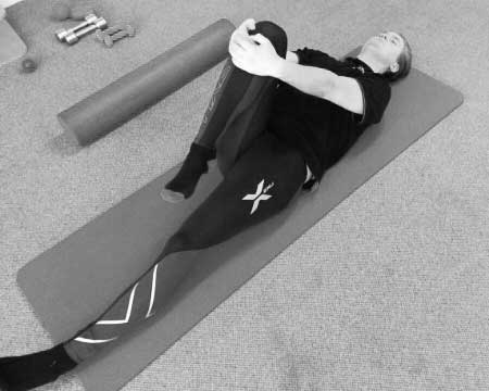
Lying face-up and bringing one knee up to your chest then pulling over to the opposite chest as we hold the stretch with our hands around our knees (i)
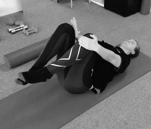
Lying face-up and placing one foot on the opposite knee and raising that knee so the hip is at 90°. Pushing the rotated leg's knee away from your head as you pull the other knee from around the back towards your chest. (ii)

(i)

(ii)
To strengthen the joint we use isometric exercises, which is a constant muscle contraction where the joint doesn’t lengthen or shorten. For all the of the steps below we hold the contraction at 50% strength of the contraction for 5 seconds repeating 10 times.
We begin by lying face up with your knees up, placing both hands with your fingers interlocked and pushing your knee into your hands then alternate.
Now have one hand under the front of the knee and the other above the knee and pushing the knees in opposite directions into the hands which are preventing the knees from moving up or down.
Repeat exercise as you alternate legs.

Next we make a fist with each hand, placing them in between the knees and pushing the knees together into the fists.

Lastly, we’re looking to push the knees outwards, so with our hands placing on the outside of the knees and pushing outwards.
An easier way to do the last exercise is a ‘clam’ which begins by lying on your side with your hips slightly bent and your knees at 90°. Keeping your feet together and your hips stable, bring your top knee up as high as you can and holding at the top for a contraction through your glutes as you return slowly to starting position. Repeat this 16 times at 4 sets, holding the contraction when the knee is at the top for 3 seconds.

Starting position

Final position
Please note these exercises are simply a guide and it's recommended to get assessed by a Richmond Rehab therapist for a more tailored exercise and treatment plan.