

Thoracic Outlet Syndrome

Thoracic outlet syndrome, commonly abbreviated to TOS, may very well be one of the most difficult and controversial diagnosis in clinical medicine.
The goal of this Blog is to breakdown this condition and to better understand what we as musculoskeletal type practitioners can do to provide the best care for patients presenting with possible symptoms of TOS. Whether that be assessing the condition, providing hands on or exercise based treatment or even knowing when best to refer on a patient that may need further investigation or even surgical intervention.
To understand the condition you first must understand the area, consisting of a group of three spaces between the clavicle, the first rib to the axilla which several important neuro-vascular structures pass. Structures of which include the brachial plexus, subclavian artery, and subclavian vein as seen in image 1 below.
Compression will primarily occur either in the inter-scalene triangle, the costo- clavicular space or the sub-coracoid space.
Inter-scalene Triangle
The first and most medial area created by the borders of the anterior scalene muscle, middle scalene and the first rib. This is where the brachial plexus and subclavian artery pass through the inter-scalene triangle.
Costco-clavicular Space
The second area is bordered by the subclavius muscle, clavicle, the first rib and anterior scalene muscle, this is where the brachial plexus, subclavian artery and subclavian vein all pass through.

Image 1
Subcoracoid Space
The pectoralis minor muscle forms the anterior border and the ribs form the posterior boundary. The brachial plexus passes through this space, and the subclavian artery and vein continue through it as the axillary artery and vein.
We also need to take into account the muscles that border these areas that the neurovascular structures pass through, as muscle hypertrophy and overuse can contribute to the compression of these structures.
Types
TOS is widely known to consist of 3 main types, which come with differing symptoms, diagnostic criteria and standard treatment strategies.

Neurogenic TOS
A presentation within the clinic where the Brachial Plexus is compressed
See image 1

Vascular TOS
A less common presentation but one that needs considerable treatment due to the compression of the subclavian artery +/or vein
Within this type, there are four sub-categories
1. Acute Thrombosis
A sudden blockage or clot
2. Chronic Stenosis
Long term narrowing
3. Intermittent obstruction without Thrombosis
Closing & opening of the vein or artery due to obstruction that isn’t a blood clot
4. Complete Obstruction
The vein or after is completely blocked & is not allowing blood to flow through

Non-specific TOS
When objective tests can’t differentiate between Vascular or Neurogenic, the presentation is termed as Non-specific.
(Povlsen, Hansson, Povlsen, 2014)
Movements
People with the shoulder flexed for majority of the work day (e.g. over head actions)
Have repeated trauma to the shoulder joint
Extended duration in compromising shoulder positions, those who exhibit abnormal posture, including positions required to play bowed instruments.
Repeated trauma to the head or neck
Postural dysfunction
Pregnancy, oedema, anatomical deviations, hypertrophied muscles, boney growths, and muscle weakness are all theorised to be contributing factors to TOS.
The most commonly presented cause of TOS appears to be caused by a whiplash motion that can result in instability at the atlantoaxial joint (C1-C2), causing the surrounding musculature such as the sternocleidomastoid and scalenes to shorten and alter their function to compensate for the laxity in the joint. This can lead to an entrapment of the brachial plexus, subclavian artery, subclavian vein, or a combination of these vessels and tissues (Levine & Rigby, 2018).
Symptoms
As mentioned previously the symptoms you may present with will indicate whether you have either definitive Neurogenic, Vascular or be a symptom of both.
Due to compression you may experience localised pain in the neck or shoulder, or alternatively in the upper arm or even referred down to the hand due to the structures that are compressed extending and branching down into the hand and fingers.
This may also illicit weakness in the corresponding limb as these nerve branches control the muscle contractions of the arm and hand.
A true symptom of Neurogenic TOS is the decrease in muscle mass of a muscle in the base of the thumb (Abductor Pollicis Brevis) or intrinsic muscles in the hand.

In regards to a Vascular specific diagnosis symptoms will usually include swelling, cyanosis (extremity turning blue) or even a decreased and difficult to read pulse (Jones et al. 2019, Povlsen et al. 2014).
Testing
So, if TOS is difficult to diagnose then how exactly do we get to the stage where we believe that is what the patient is presenting with and to then provide a gold standard treatment?
Well if you have seen a Physiotherapist or a Myotherapist at Richmond Rehab or even elsewhere you will be very familiar with how many questions we ask, to get a better understanding of your symptoms and to try and tease out differentiating symptoms to narrow down our testing.
By this stage of a session if you have mentioned any of the previous symptoms your therapist will likely undergo the following objective tests to confirm or count out TOS.
Adsons Test
A manoeuvre where the therapist will find the patient's radial pulse down near their wrist and then proceed to move the patient's arm upright in an arc compressing the subclavian artery. The therapist will gauge whether the patient's pulse diminishes and then compare to the opposing non-symptomatic side.
Roos Test (Elevated Arm Stress Test) - Across
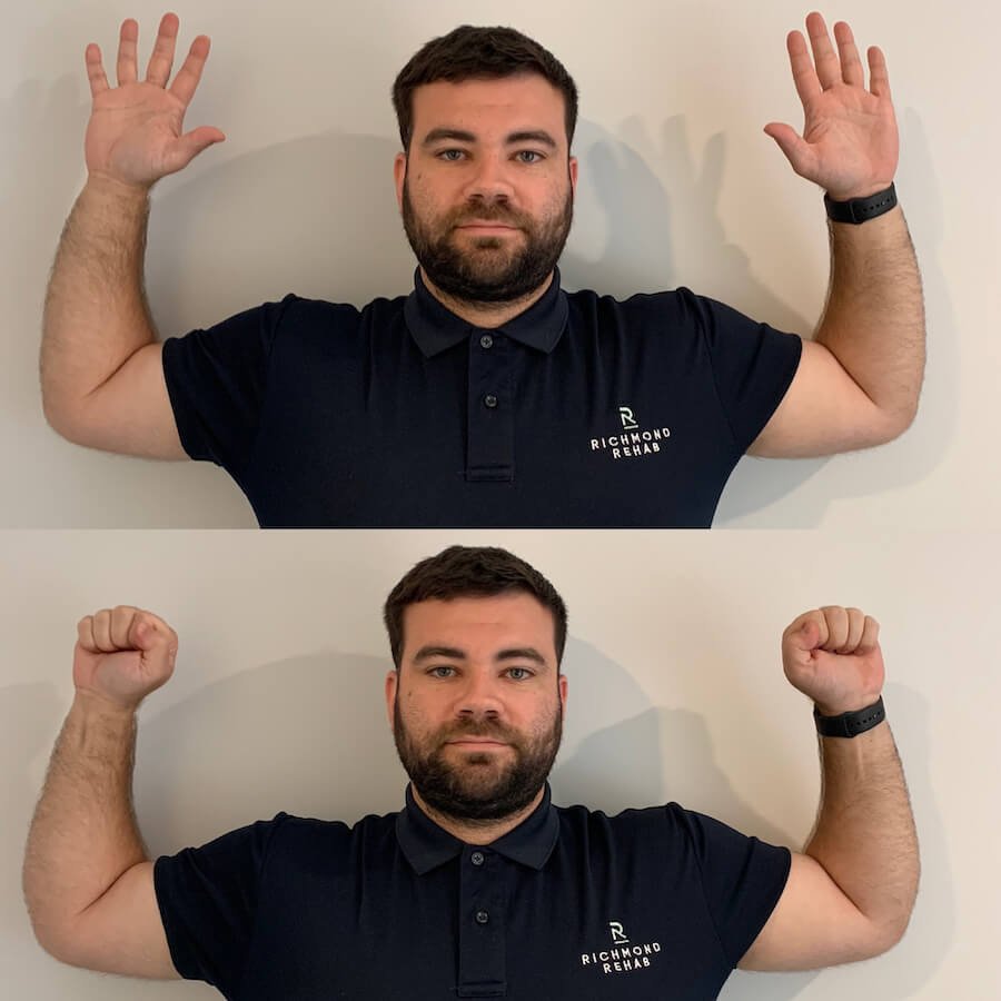
This is a quite easy one for the patient to perform and works well for inter-therapist reliability. The patient stands in with both shoulders and elbows at 90 degrees and externally rotated in the “surrender position”. They will then squeeze their hands for up to 3 minutes or until symptoms occur.
Adson’s test has a 92% sensitivity and Roos a 98%, meaning it has a 92% and 98% chance respectively of identifying a condition within the tissue being tested. However where this gets difficult is the specificity is quite low, documented to be below 80% even as low as 18%, meaning that althought symptoms are present we as therapist cannot definitively say that the patient has TOS. Where this changes however is when combined and both showing positive results the specificity increases to approximately 82% (Jones et al. 2019, Sadeghi-Azandariyani et al. 2009)
Imaging
One of the greatest tools we have as therapists is the referral network, specifically knowing who or when to refer someone on for a condition or set of symptoms that either may need further investigation or more importantly a condition that we are unable to treat.
TOS is a condition that is important in its diagnosis of true neurogenic or vascular types.
Neurogenic
When suspecting neurogenic TOS symptoms a nerve conduction study may be referred by your doctor, neurosurgeon or neurologist, as well as an EMG (Electromyography) to test the nerves that run down your arm branching off the brachial plexus that may be compressed.(Povlsen et al. 2014)
Vascular
If a vascular type of TOS is suspected or even to differentiate a non-specific or disputed type of TOS then imaging is essential.

The three most common tests you will be referred for are either a Chest Radiograph (X-ray), MRI or a CT scan, alternatively an ultrasound has high sensitivity and specificity. These are the preferred non-invasive techniques, however if the patient requires urgent care for an acute vascular TOS then an angiograph may be required (Raptis et al. 2016, Jones et al. 2014).
If you believe you may have symptoms of vascular TOS then a consultation with your GP would be recommended.
Treatment Strategies
The main goal for treating TOS would be to treat any muscle imbalance, decrease muscle tension and increase muscle length of shortened structures improving symptoms of nerve compression.
A range of Myotherapy techniques can be used to assist relaxing the muscles surrounding the area and assist restoring postural faults that could have been contributing to compression of the thoracic outlet. Hands-on treatment may include massage, myofascial release, trigger point therapy and post-isometric relaxation stretching techniques. These treatment techniques are most effective when combined with correct stretching and strengthening exercises.
Co-management of TOS is also fundamental to recovery. A myotherapist can work alongside other allied health or medical practitioners to help manage thoracic outlet syndrome. Management would include improving upper body posture through education, implementing strength exercises, changing movement patterns, sleeping position or recreational activities.
Management
Ok, now you have been to your therapist and you have a working clinical diagnosis of TOS and you’re up to the stage where you need to do something about it!
The options are Operative or Non-operative

One we are well trained to provide and the other unfortunately will have to be done by a trained surgeon...
Treatment and management strategies of neurogenic TOS are recommended for a minimum of four months prior to engaging in surgical intervention.
Rehab is the initial recommended management and should include Physiotherapy based exercises for postural mechanics to decrease the soft tissue compression. Exercise prescription alone is difficult to achieve the desired results and will often work well with activity modification advice and education from your physiotherapist and myotherapist and relaxation techniques mentioned previously best administered by a myotherapist or remedial massage therapist (Jones et al. 2019).
Additional treatment strategies like pharmacological treatments will assist in the improvement of pain symptoms and neurological symptoms and should be discussed with your GP.
With regards to a vascular type of TOS, a surgical intervention is at times undertaken prior to musculoskeletal rehab with a Physio. Post-surgical management will likely involve a comparable rehab strategy as a non-operative Neurogenic TOS (Povlesen et al. 2014).
This condition like a lot of other conditions is not a cookie cutter method, meaning that the treatment largely is based on symptoms and presentation rather than a blanket treatment for all TOS patients. The best thing you can do for treatment of this condition is have a therapist assess you and plan out the treatment strategy or as mentioned previously refer on for further investigative assessment.